22 Post-Lab for Axial Skeleton
Gillian Backus; Heidi W. Wangerin; and Paula Rodgers
Post-Lab: Axial Skeleton
Name: ________________________
Lab Checkout:
When you finish the lab, please clean up your lab space and put away your materials neatly in the tray. Once you have thoroughly cleaned, washed, and dried your lab table, please get your instructor’s initials to check-out of lab.
- Lab bench clean, washed, and dried
- Materials put away properly and organized in trays
- Microscope properly put away
Lab completed (% completed = ______ %) Instructor initials: ______________
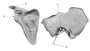
Activity 1: Bone Marking Terminology
1. Use the bone marking chart from Activity 1 to select an appropriate bone marking term to best describe the structures labeled A-D. (ideas: condyle, tubercle, crest, spine)
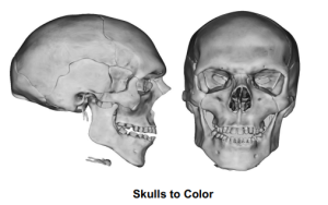
Activity 2: Bones and Bone Markings of the Skull
2. Color the bullet point (or term) and the corresponding bone on the skull image.
-
- ethmoid bone
- frontal bone
- hyoid bone
- inferior nasal
- lacrimal bone
- mandible bone
- maxillae bone
- nasal bone
- occipital bone
- palatine bone
- parietal bone
- sphenoid bone
- temporal bone
- vomer bone
- zygomatic bone
3. Highlight the bullet point (or term) and color in the corresponding sutures of the skull with the same color:
-
- coronal suture
- lambdoid suture
- sagittal suture
- squamous suture
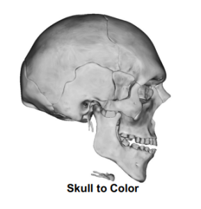
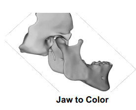
4. Color the bullet point (or term) and the corresponding surface of the bone marking on the mandible and temporal bone with the same color.
-
- coronoid process
- mandibular condyle
- mandibular foramina
- mandibular notch
- mandibular ramus
- mental foramen
- external acoustic meatus
- mandibular fossa
- mastoid process
- styloid process
- zygomatic process
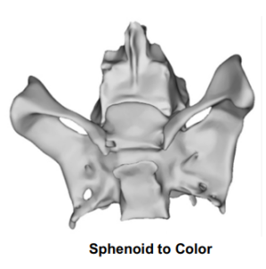
5. Color the bullet point (or term) and the corresponding surface of the bone marking on the sphenoid and ethmoid bone in the same color:
-
- greater wings
- lesser wings
- sella turcica
- cribiform plate
- crista galli
6. Color the bullet point (or term) and the corresponding fontanels on the fetal skull in the same color (A = anterior, P = posterior, M = medial, L = lateral).
-
- anterior fontanel
- posterior fontanel
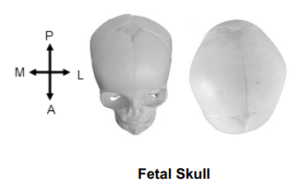
7. Provide at least two functional reasons babies have fontanels.
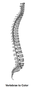
Activity 3: Bones and Bone Markings of the Vertebral Column
8. Color the bullet point (or term) and the corresponding vertebrae in the same color.
-
- cervical vertebrae
- coccyx
- lumbar vertebrae
- sacrum
- thoracic vertebrae
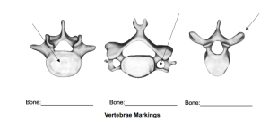
9. Identify the type of vertebrae. Label the parts indicated at the arrows.
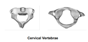
10. How are the atlas and axis different from the typical cervical vertebrae? Label the unique bone markings in the images below.
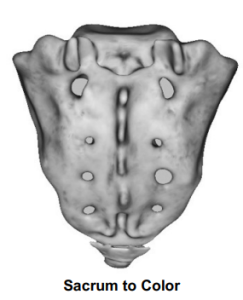
11. Color the bullet point (or term) and in the same color the corresponding area on the sacrum.
-
- sacral canal
- sacral foramen
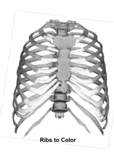
Activity 4: Bone and Bone Markings of the Thoracic Cage
12. Describe the difference between true ribs and false ribs.
13. Color the bullet point (or term) and the corresponding bones and bone markings of the ribs and sternum in the same color.
-
- body
- false ribs
- manubrium
- true ribs
- xiphoid process