
10 Muscles Lab
"No knowledge can be more satisfactory to a man than that of his frame, its parts, their functions and actions." -Thomas Jefferson
Gillian Backus; Heidi W. Wangerin; and Paula Rodgers
Objectives
- Identify the muscles of the body on a model or image.
- Identify a muscle on the body in anatomical position as being anterior, posterior, medial, lateral, superficial and/or deep.
- Identify the major actions of each muscle listed using terms like flexor, extensor, adductor, abductor, rotator etc.
- Identify the origin and insertion points of selected muscles (with an asterisk *) using terms from your axial and appendicular lab.
- Define, identify, and label a tendon, ligament, and aponeurosis.
- Given the origin and insertion of selected muscles (with an asterisk *), predict the action of a muscle.
Terminology Checklist
Muscles of Facial Expression
|
Muscles of Respiration
|
Muscles of Chewing
|
Muscles of the Abdominal Wall
|
Muscles of the Head, Neck, and Vertebral Column
|
Muscles of the Pectoral Girdle and Arm (Humerus)
|
Muscles of the Forearm (Radius & Ulna)
|
Muscles of the Tibia and Fibula
|
Muscles of the Carpus and Hand
|
Muscles of the Ankle and Foot
|
Muscles of the Hip and Thigh (Femur)
|
Outline of Lab
Case Study: “Too Tired to Stand”
Activity 1: Muscle Actions
Activity 2: Muscles of the Axial Skeleton
Activity 3: Muscles of the Appendicular Skeleton
Putting it all Together
Case Study: “Too Tired to Stand”
You are working as a nurse’s assistant in a pediatrician’s office. As you bring the next patient, a 5-year-old boy named Jose, back to the exam room, he trips, and you catch him. His parents comment that he falls frequently. You observe how he moves and notice that he walks on his toes and uses his arms for balance. You notice that his calves appear larger than the average child of his age. When you get to the exam room, he is mildly out of breath. After questioning his parents, you learn that Jose has trouble in school and suspect he has a learning disability. The parents are concerned that Jose has trouble running and jumping. He also frequently complains of muscle pain. When you question Jose about the pain, he comments that today his shoulder is sore. You ask him to show you how he ties his shoes. After he is done, he places his hands on his ankles and “walks” them up to his shin, knee and then thigh to stand upright. (This method of standing up is called “Gower’s sign”.) The pediatrician will want to see that you include all the muscles that might be affected in your notes. It might be wise to review the major muscles of the body!
Activity 1: Muscle Actions
Materials:
- none
Background:
There are over 600 muscles in the human body. Learning their names, locations and actions can be intimidating. In this lab we will focus on a select subset of muscles that contribute to major body movements. Skeletal muscle usually attaches to two different bones, one called the origin and the other at the insertion, and typically spans a joint. Muscles connect to the bone by a cord of dense connective tissue, called a tendon. When the muscle contracts, it gets shorter as it pulls one bone (insertion) toward the other bone (origin). Thus, in most cases the origin is stationary while the insertion moves toward the origin.
Exceptions to the rule
Some muscles insert not on bone, but on fascia, tendons, or even collagen fibers of the dermis. An aponeurosis is a broad sheet of dense connective tissue (tendon) that can serve as a site for muscle attachment. Instead of using the terms origin and insertion, anatomy books may refer to the proximal and distal sites or superior and inferior attachment sites for a muscle.
Muscle Actions
While we may think of each muscle as having a singular function, many muscles have multiple actions. Moreover, many muscles act together to carry out the same function. The actions of muscles can be grouped into four categories: prime mover (agonist), synergist, antagonist, and fixator. The prime mover (agonist) is a muscle that produces the most force during a particular movement. A synergist is a muscle that assists the prime mover (agonist) during the movement. One or more synergistic muscles allow for stronger contractions than can be produced by a single muscle on its own. Many synergistic muscles do not simply perform the same action as the prime mover but allow for a slightly different movement. For example, a synergist might assist the prime mover with flexion, creating greater flexion, but also contribute to rotation of that joint.
Every movement must have an opposing action; that is the job of the antagonist. Just like a villain or antagonist in a movie opposing a hero character, the antagonist opposes the action of the prime mover (agonist). For example: when the prime mover of the elbow flexes, the antagonist is relaxed. Similarly, when the prime mover of the elbow contracts to extend the elbow, the flexor must relax.
Lastly, a fixator, “fixes”’ a bone in a position; a fixator prevents a bone from moving and instead holds it in a static position. Fixator muscles maintain posture, as in the muscles of the back, abdomen, and neck.
A joint is formed where two bones meet. As a muscle contracts, it pulls on the bone to which it is attached and causes the joint to move. Specific movements occur at the articulating surface of all synovial joints. The type of movement allowed at each synovial joint depends on the shape of the articulating bones and the direction of the muscle fibers causing contraction. Actions are classified as angular, gliding, and special movements. For this lab we will focus on the angular and special movements. These actions come in opposing pairs. Angular movements are the most common actions. Such movements increase or decrease the angle between two bones and include flexion, extension, adduction, abduction, medial and lateral rotation. Special movements include supination, pronation, dorsiflexion, plantarflexion, inversion, eversion, protraction retraction, and elevation and depression. (Joints will be explored in more depth in a separate lab.) (Table 1)
Table 1: Classification of muscle actions and their description.
| Angular or Special | Movement | Description | Opposing movement |
| A | Flexion | Decreases the angle between bones; brings bones closer together. Movement in an anterior-posterior plane | Extension |
| A | Lateral flexion to the right | The vertebral column moves in the lateral direction along the coronal plane | Lateral flexion to the left |
| A | Abduction | Lateral movement of the body part away from midline | Adduction |
| S | Pronation | Rotation of the forearm where the palm is turned posteriorly (radius rotates over ulna) | Supination |
| A | Dorsiflexion | Ankle joint bends so that the dorsum (superior surface of the foot) moves toward the leg | Plantar flexion |
| A | Inversion | Movement at the ankle joint that turns the sole of the foot medially or inward | Eversion |
| A | Medial or internal rotation | Moving the anterior surface of the limb toward the midline of the body | Lateral or external rotation |
| S | Protraction | Movement of the scapula or mandible in the anterior (forward) direction. | Retraction |
| S | Elevation | Movement of the scapula or mandible in the superior (upward) direction. | Depression |
Interpreting muscle names
While the names of skeletal muscle names might seem complicated, there are clues and patterns to their names that you can learn. Some muscles are named for their location in the body. For example, the intercostal muscles are found between the costal spaces of the ribs. The intermedius muscles are located between two other muscles. Some muscles are named for their unique shapes and sizes. For example, the trapezius muscle has a trapezoidal shape. Names such as maximus (largest), minimus (smallest), longus (long), and brevis (short) give a hint as to the relative size of the muscle. Muscle fibers run in different directions; some names indicate the direction of the fibers. For example, the term rectus means straight, transversus means horizontal., and oblique indicate fibers running at an angle. Some muscles use the origin and/or insertion in their name. The sternocleidomastoid muscle is named for both its origins (sternum and clavicle) and insertion (mastoid process). In addition, the number of “heads” or origins can be used in a name as in the quadriceps muscle which has four heads or the two-headed biceps muscle of the arm. Muscle actions may also be used as part of the name. The extensor digitorum, for example, extends the digits of the hand while the flexor digitorum superficialis flexes the digits of the hand.
Key for muscle names
Location
|
Direction of fibers
|
Number of heads
|
|
Shape
|
Size of the muscle
|
Learning muscles
1. Look at the name of the muscle
- Many muscles are named based on their shape, location, action, fiber direction, or muscle attachment sites.
- Think about what hints the muscle name might give you.
2. When possible, palpate the muscle on yourself. Contract the muscle to feel it bulge and see what bones move.
3. Look at an articulated skeleton to examine how the origin and insertion dictate the movement.
4. Group muscles together based on action. Typically, muscles with similar actions are located close to each other.
Procedure:
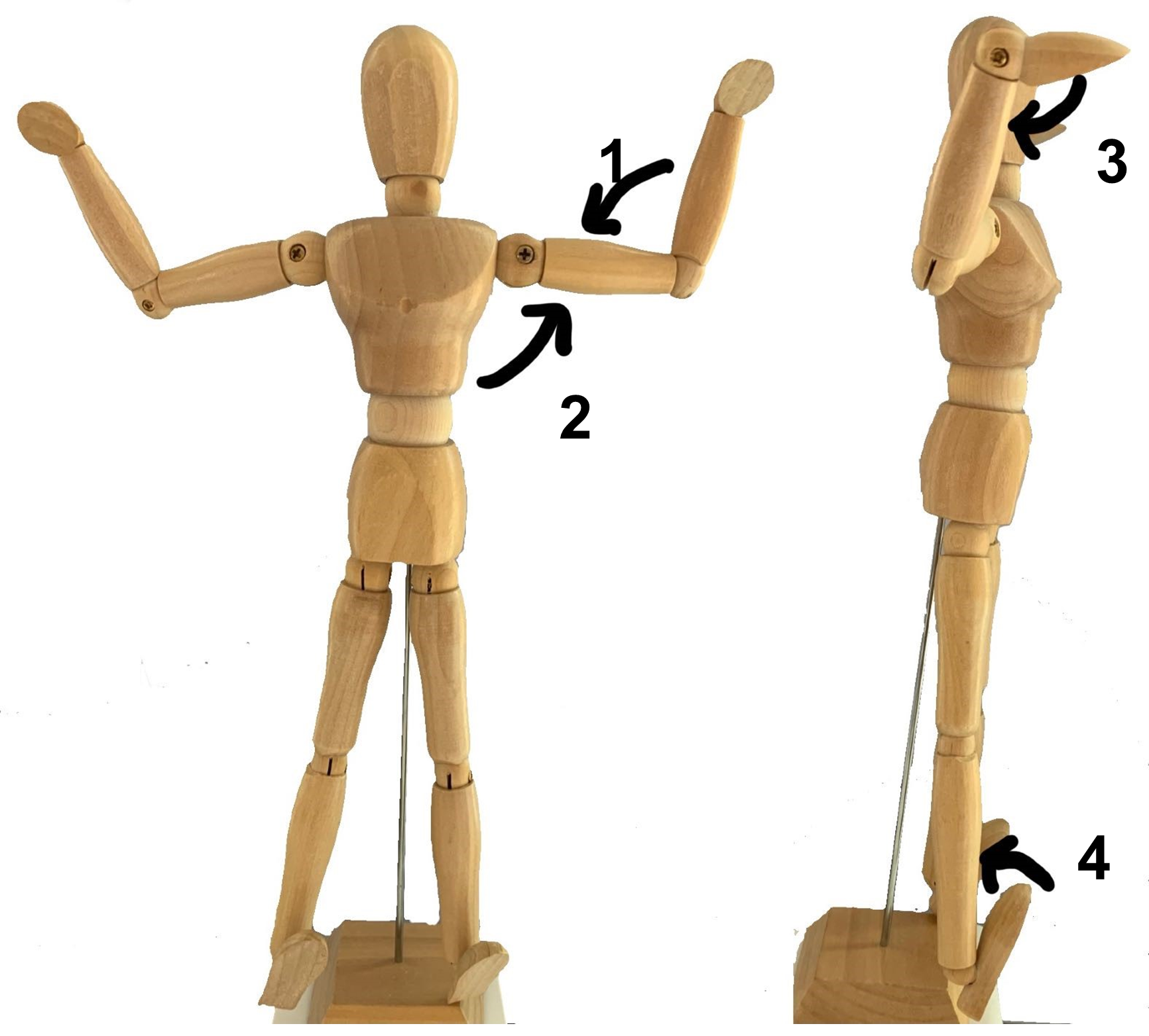
1. Use the images of angular movements below and identify the movement (use Table 1 as a guide).
| Arrow | Movement |
| 1 | |
| 2 | |
| 3 | |
| 4 |
Joint Movements
2. Look at the images below and draw an arrow for the direction of muscle contraction and name the angular movement using the terms from Table 1.
Muscle Action Predictions
Movement: ______________________ ______________________ ______________________ ______________________
Activity 2: Muscles of the Axial Skeleton
Materials:
- laminated numbers 1-5
- laminated terminology labels with sticky tack
- model of body muscles
- model of head muscles
Background:
In the patient’s notes, you see new results from a recently performed genetic test. Jose has the gene for a genetic disease called Duchenne type muscular dystrophy. It is a progressive disease caused by a defective gene for dystrophin (a protein in muscle). The condition is more common in boys than girls, as it is carried on the X chromosome. As the disease progresses, patients develop severe muscle weakness, including the diaphragm muscle and the intercostal muscles which makes it difficult to breathe. You notice that Jose has a protruding belly despite not being overweight. He is slumped in the chair, and has difficulty catching his breath. Silently, you review the muscles that are important for breathing, supporting the abdomen, and holding up the frame of the body.
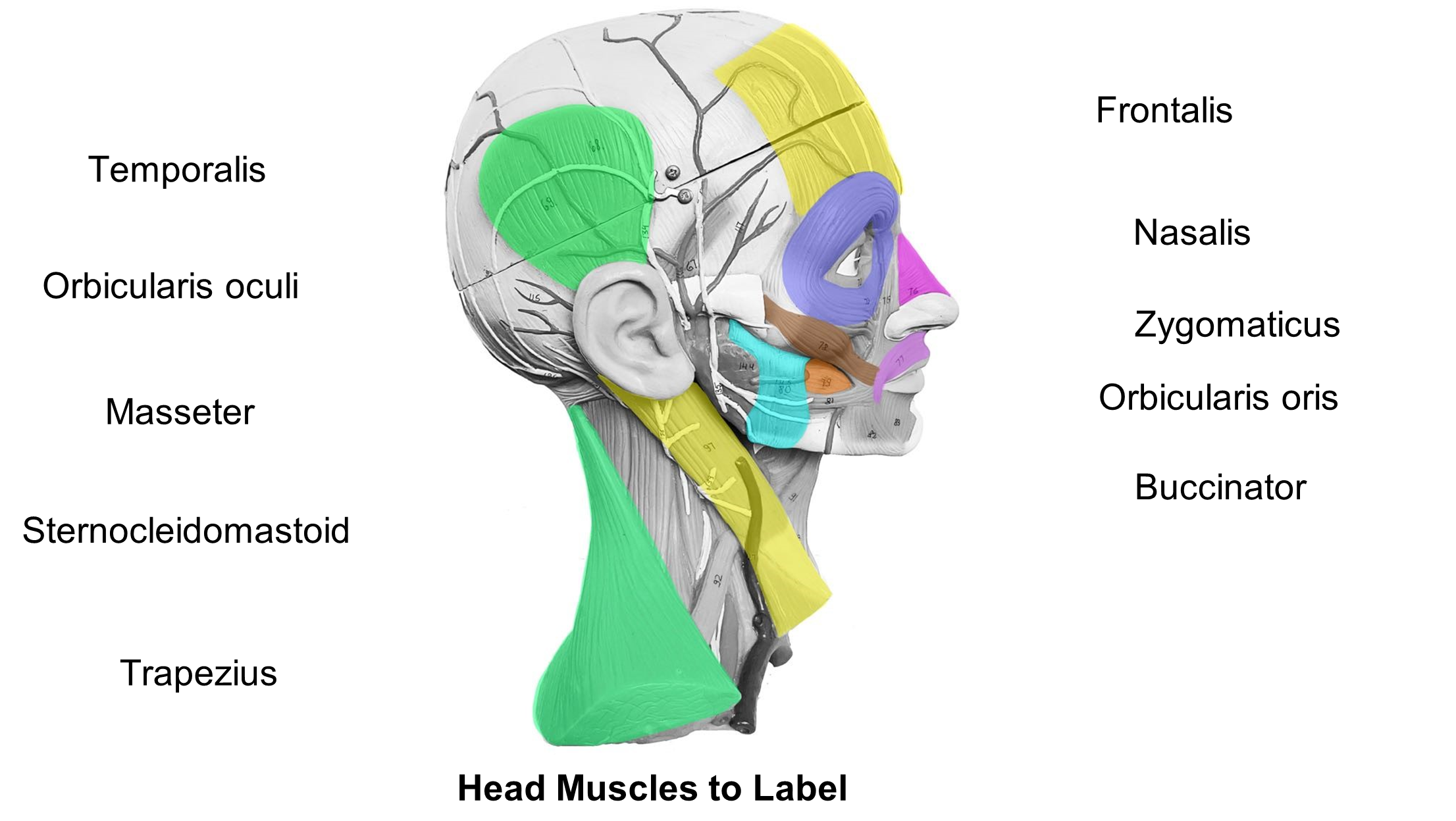
Muscles of facial expression (Table 2, Figure 1)
Humans are social animals that communicate not only verbally, but also through facial expressions. Every facial expression is governed by numerous muscles contracting simultaneously. Most of the muscles of the face are innervated by cranial nerve 7, the facial nerve. This nerve controls many facial expressions. Indeed, when this nerve is injured, facial paralysis often occurs, causing distinctive facial sag on the affected side. To create facial expression many muscles of the face insert into the connective fibers of the dermis. This allows the skin to move as the muscles contract.
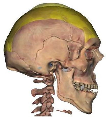
The occipitofrontalis (epicranius) is frequently divided into two parts, the frontalis and occipitalis, connected by an aponeurosis, a large band of connective tissue. The frontalis is superficial to the frontal bone and the occipitalis is superficial to the occipital bone.
Muscle names like the orbicularis oris and orbicularis oculi give you hints of shape and location in their name. Orb means circle. Oculi and oris refer to eye and mouth respectively. Look for round muscles surrounding the mouth and the eye respectively The nasalis is the muscle of the nose. The zygomaticus muscle originates on the zygomatic bone. The platysma is a large, sheetlike muscle that can be seen in the anterior neck area. It is responsible for the “jaw-dropping” expression and tightening the neck.
Table 2: Muscles of facial expression with their origin(s), insertion(s), action(s) and innervation.
| Muscle | Image | Action | Origin | Insertion | Innervation |
| Epicranius (occipitofrontalis) Frontalis & Occipitalis |
 |
frontalis: raises eyebrows occipitalis: pulls scalp posteriorly | pulls the corners of the mouth laterally and superiorly (for smiling) | frontalis: skin of eyebrows occipitalis: epicranial aponeurosis | Facial nerve |
| Nasalis | 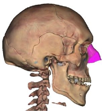 |
elevates corners of nostrils (flares the nose) | maxilla and cartilage of nose | dorsal side of nose | Facial nerve |
| Orbicularis oculi |
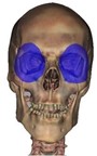 |
closes eye (blinking and squinting) | orbital portions of the frontal bone and maxilla | skin of orbital area and eyelids | Facial nerve |
| Orbiularis oris |
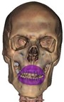 |
compresses and purses lips | maxilla and mandible | connective tissue of the lips | Facial nerve |
| Platysma |  |
depresses the lower lip, depresses the mandible | connective tissue of deltoid and pectoralis muscles | mandible | Facial nerve |
| Zygomaticus major |
 |
pulls the corners of the mouth laterally and superiorly (for smiling) | zygomatic bone | corner of mouth | Facial nerve |
Muscles of chewing (Figures 1, 2, and Table 3)
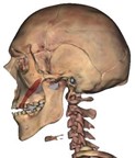
The fibers of the buccinator run transversely, and when contracted, compresses the cheek (Figure 1). Buccinator means trumpeter. A trumpeter must compress the cheeks to expel air from the mouth. The masseter muscle also hints at its action-mastication (chewing). The lateral pterygoid is deep to the buccinator and masseter, it also aids in chewing. The temporalis muscle is located superficial to the temporal bone (Figure 3).
Figure 1: Muscles of the head and face involved in facial expression and chewing.
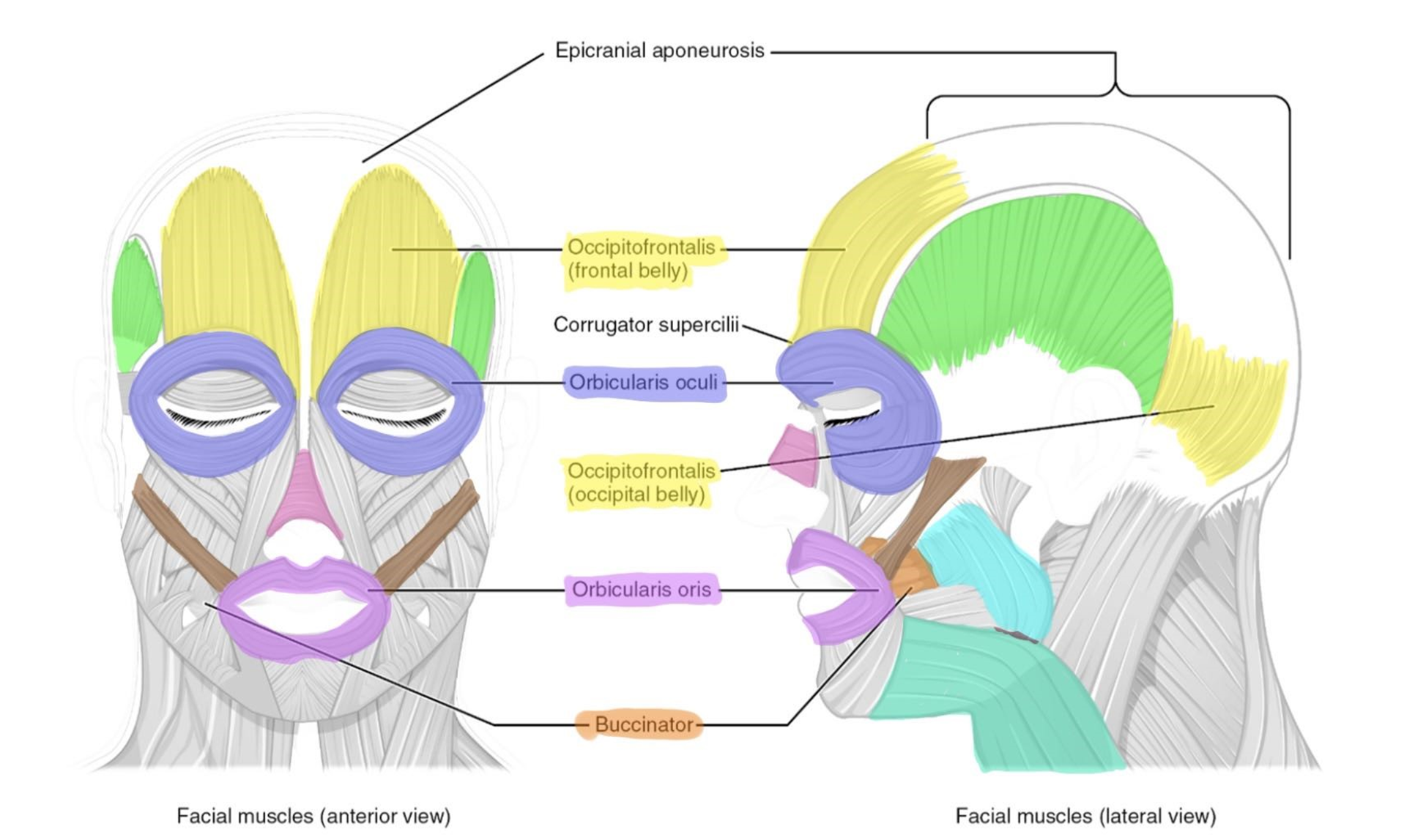
Figure 2: The superficial (left) and deep (right) muscles involved in chewing.
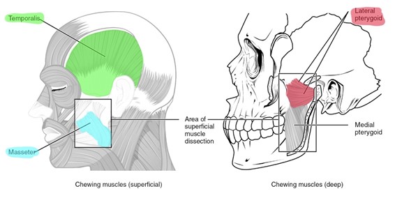
Table 3: The action(s), origin(s), insertion(s) and innervation of the muscles involved in chewing.
| Muscle | Image | Action | Origin | Insertion | Innervation |
| Buccinator |  |
compresses cheek, pulls checks in (sucking movement) | maxilla and mandible | connective tissue of cheeks and obicularis oris | Facial nerve |
| Lateral pterygoid |
 |
protracts and depresses the mandible | greater wing of the sphenoid | mandible | Mandibular nerve |
| Masseter | 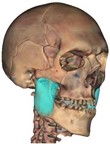 |
closes (elevates) the jaw | zygomatic arch | angle and ramus of mandible | Mandibular nerve |
| Temporalis | 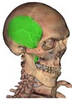 |
elevates and retracts the mandible (closes the jaw) | lateral surface of skull | coronoid process of the mandible | Mandibular nerve |
Figure 3: Muscles of the neck and back.
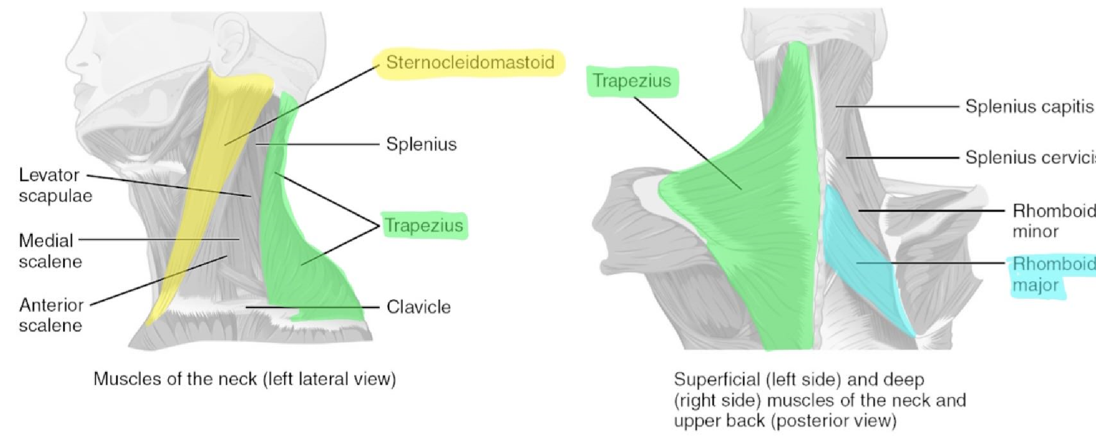
Muscles of the head, neck, and vertebral column (Figures 3, 4 and Table 4)
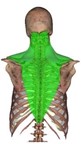
The sternocleidomastoid muscle tells you the origin and insertion in the name. The sternum and clavicle are the origins, and the insertion is the mastoid process of the temporal bone. Since the muscle starts medially and ends laterally, this muscle causes a twisting (rotational) movement of the head.
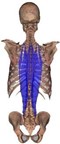
The erector spinae group is group of muscles responsible for extension of the vertebral column. They help in flexion, lateral flexion, and rotation of the vertebral column. The three main muscles are the iliocostalis (lateral), the longissimus (middle), and spinalis (medial) (Figure 4). Other back muscles will be discussed when shoulder and arm movements are discussed (See pectoral girdle and scapula).
Figure 4: The erector spinae muscle group.
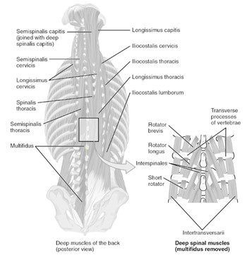
Table 4: Muscles of the neck and vertebral column.
| Muscle | Image | Action | Origin | Insertion | Innervation |
| Erector spinae *Spinalis (medial) *Longissiumus (intermediate) *Iliocostalis (lateral) |
 |
extend and laterally flex the vertebral column | vertebrae | vertebrae | Posterior rami of spinal nerves |
| Sternocleidomastoid | 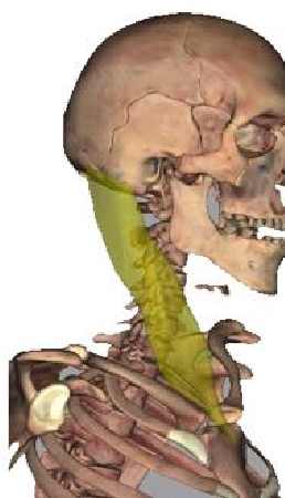 |
flexes head, laterally rotates head | manubrium of sternum and medial portion of clavicle | mastoid process of temporal bone | Accessory nerve |
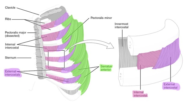
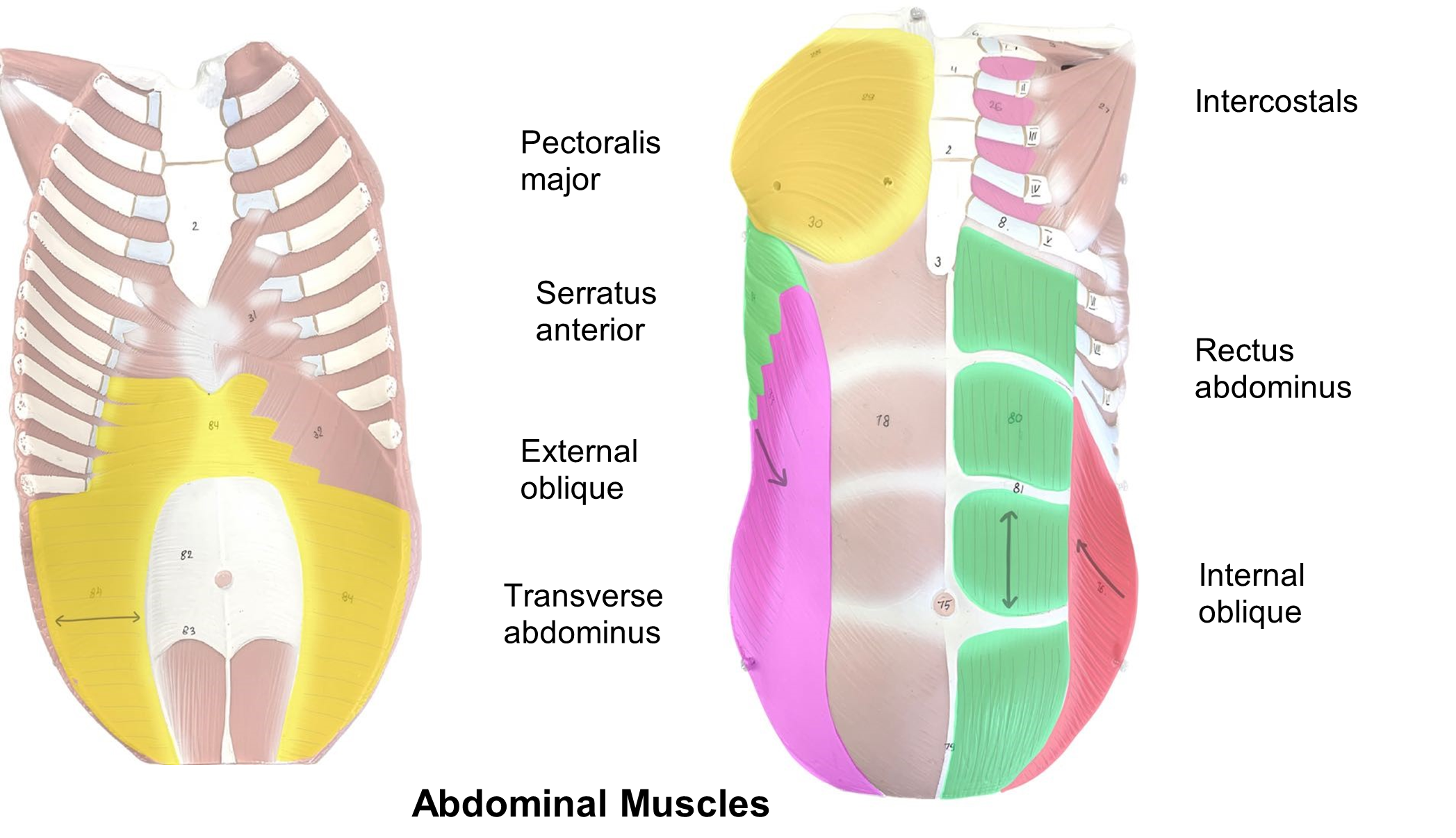
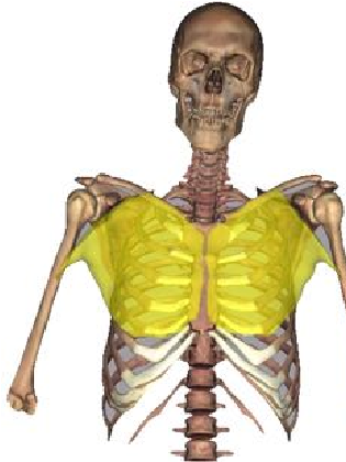
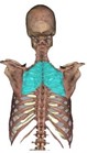
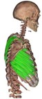
Muscles of Respiration (Figure 5, Table 5)
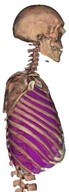
Most thoracic muscles assist in breathing, inhalation, and exhalation. The diaphragm is a large muscle that separates the thoracic and abdominal cavities. The muscle is dome-shaped at rest and flattens during contraction pulling the ribs and increasing the size of the thoracic cavity for inhalation. Relaxation of the diaphragm assists in exhalation. The external intercostal muscles are located outside of each rib and assist in inhalation while the internal intercostals are located deep to each rib and contract the rib cage during forced exhalation. The fibers of the external and internal intercostals run opposite to each other. When looking at the patient’s right side, the external fibers run like a forward slash (\) and the external intercostals run like a backwards slash (/). Together the fibers create an “X” pattern.
Table 5: Muscles of respiration, their action(s), origin(s), insertion(s) and innervation.
| Muscle | Image | Action | Origin | Insertion | Innervation |
| External Intercostals |
 |
elevates the rib cage, assists in inspiration | inferior edge of superior rib | superior edge of inferior rib | Intercostal nerves |
| Internal Intercostals |
depresses the rib cage, assists in forced expiration | lateral edge of costal groove of inferior rib | superior rib, deep to the external intercostal attachment | Intercostal nerves | |
| Diaphragm | 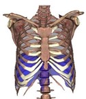 |
flattens the thoracic cavity for inspiration (inhalation) | xyphoid process of sternum, lower ribs, costal cartilages, and upper lumbar vertebrae | central tendon of diaphragm | Phrenic nerve |
Figure 5: Muscles involved in breathing (ventilation).
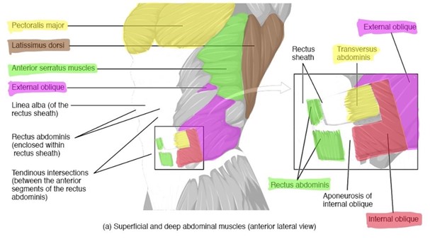
Muscles of the abdominal wall (Figure 6, Table 6)
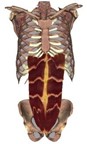
There are four pairs of muscles that make up the main abdominal muscles. Three of the muscles are located anterolaterally and are layered on top of each other. The external oblique is the most superficial and can be identified based on the direction of the fibers. Place your hands in your pockets and the direction of your fingers represents the fibers of the external oblique. If you cross your arms with your fingers on your shoulders this would represent the direction of fibers of the internal oblique. The deepest muscle is the transversus abdominis and as the name implies the fibers run transversely (horizontally) across the abdomen. The last muscle is the rectus abdominis. The term rectus means straight. The fibers of the rectus abdominis run straight up and down the abdomen and create the “6-pack” look with three transverse bands of collagen fibers between each section of muscle. The linea alba (white line) runs down the midsagittal plane of the abdomen. It forms the rectus sheath, the attachment site for some abdominal muscles.
Table 6: Muscles of the abdominal wall and their action(s), origin(s), insertion(s)
and innervation.
| Muscle | Image | Action | Origin | Insertion | Innervation |
| External oblique |
 |
flexes and laterally flexes the trunk; compresses abdomen | outer surface of Inferior eight ribs | lateral iliac crest, pubic tubercle, and linea alba | Thoracic nerves |
| Internal oblique |
 |
flexes and laterally flexes the trunk; compresses abdomen | iliac crest | inferior border of lower four ribs, linea alba, and pubic bone | Thoracic nerves |
| Rectus abdominis |
flexes trunk; compresses abdominal cavity | superior part of pubic bone | costal cartilages of lower rib bones and xiphoid process | Thoracic nerves | |
| Transverse abdominis |
 |
compresses abdominal cavity | inferior costal cartilage of lower 6 ribs, medial iliac crest, and fascia | linea alba and pubic crest | Thoracic nerves |
Figure 6: Muscle of the abdominal wall.
Procedure:
1. Divide into pairs. One group label the muscles on the head model while the other labels the muscles of the abdomen and back. As you identify the muscles, label them with the laminated muscle stickers.
2. As you identify the muscles, label them with the laminated muscles terms and label the corresponding images by drawing a line between the muscle name and the muscle.
Activity 3: Muscles of the Appendicular Skeleton
Materials:
- laminated terminology labels with sticky tack
- model of full body muscles
- model of muscle arm
- model of muscle leg
Background:
Today the doctor will perform a muscle biopsy from the rectus femoris of Jose’s leg to confirm the diagnosis. Children affected with Duchenne muscular dystrophy often are in a wheelchair by age 12-15. Their skeletal muscles are too weak to move the arms and are not strong enough to support the body to walk. You grab the supplies to prep for the muscle biopsy and review in your head precisely where the rectus femoris is located. The femoris must mean it is over the femur and rectus means straight. You hope it is the straight muscle over the femur....
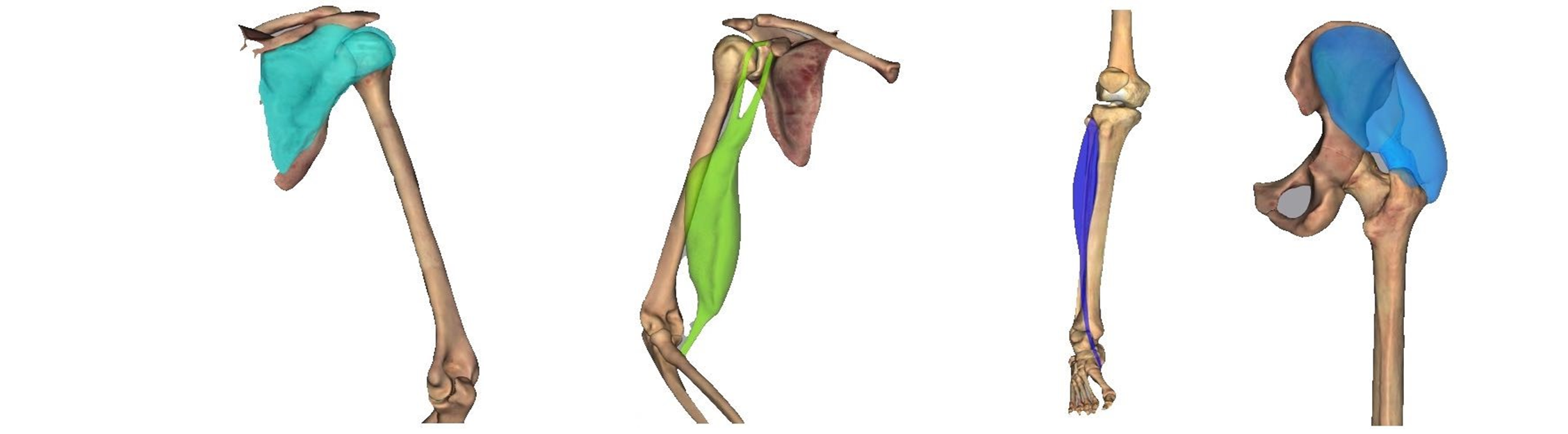
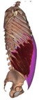
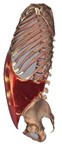
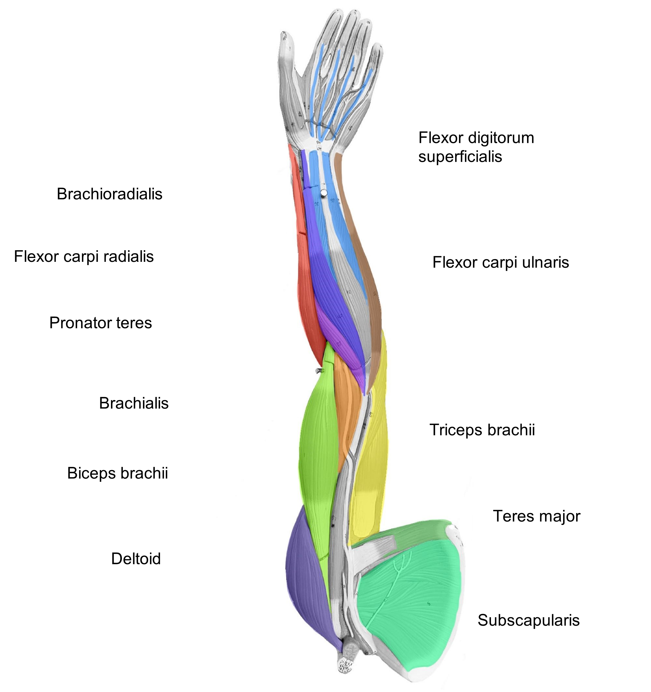
Muscles of the pectoral girdle and arm (humerus)(Figures 7, 8, Tables 7 and 8)
The muscles of the scapular region perform several movements such as elevation (superior movement of scapula), depression (inferior movement of scapula), protraction or abduction (lateral and anterior movement of scapula), retraction or adduction (medial and posterior movement of scapula), and rotation (upward and downward rotation of the scapula). These muscles include the pectoralis major, the serratus anterior, and the muscles of the rotator cuff.
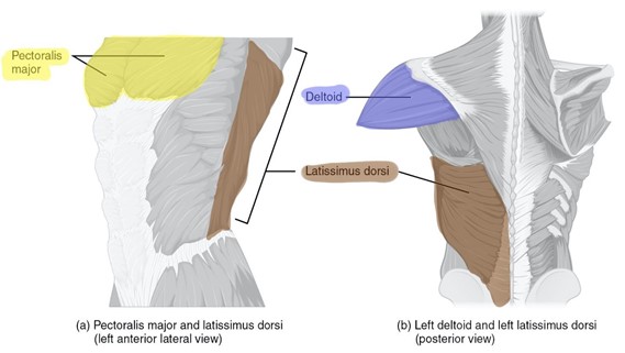
The trapezius muscle is a diamond or trapezoid shaped muscle found on the posterior head and neck. The Rhomboid (major) is found in the middle of the trapezius muscle on the dorsal surface. It is responsible for pulling or retracting the scapula. The pectoralis major is a thick, fan shaped muscle that covers the superior portion of the superficial thorax. Muscles that originate posterior to the shoulder tend to extend the arm like the latissimus dorsi and teres major. The latissimus dorsi is a large, triangular muscle found on the dorsal surface. The teres major can be found originating on the posteroinferior scapula inserting into the humerus. The serratus anterior looks like a serrated knife blade and can be found on the lateral anterior surface of the thorax.
The deltoid sits on the shoulder like a shoulder pad and is triangular shaped. The point of the triangle inserts onto the humerus between the biceps brachii and brachialis. The tendons of four muscles circle the shoulder joint forming the rotator cuff. The rotator cuff muscles include the subscapularis, located on the anterior surface of the scapula, the supraspinatus found superior to the spine of the posterior surface of the scapula, and the infraspinatus which is located inferior to the spine of the scapula. The teres minor is the fourth muscle of the rotator cuff group.
Table 7: Muscles of the pectoral girdle and arm and their action(s), origin(s), insertion(s) and innervation.
| Muscle | Image | Action | Origin | Insertion | Innervation |
| Deltoid |  |
arm abduction, secondarily flexes and extends arm | acromion and spine of scapula; acromial end of clavicle | deltoid tuberosity of humerus | Axillary nerve |
| Infraspinatus | 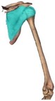 |
adducts and rotate arm laterally | infraspinous fossa of scapula | greater tubercle of humerus | Subscapular nerve |
| Latissimus dorsi |
 |
adducts, extends, and medially rotates the arm | iliac crest, spinous process of thoracic and lumbar vertebrae | humerus | Thoracodor sal nerve |
| Pectoralis Major |
 |
flexes, adducts, and medially rotates humerus | medial scapula and lateral sternum | lateral part of humerus | Medial and lateral pectoral nerve |
| Rhomboid major |
 |
retracts scapula | spinous process of T2-T5 | medial border of scapula | Dorsal scapular nerve |
| Serratus anterior |
 |
primary for protraction of scapula, and assists in rotation of scapula | ribs 1-9 | medial border of scapula | Thoracic nerves |
| Subscapularis | 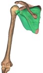 |
rotates arm medially | subscapular fossa of scapula | lesser tubercle of humerus | Subscapular nerve |
| Supraspinatus |  |
arm abduction | supraspinous fossa of scapula | greater tubercle of humerus | Subscapular nerve |
| Teres major | 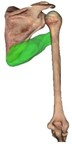 |
adducts, extends, and rotates arm medially | posterior, inferior portion of scapula | lesser tubercle and intertubercular groove of humerus | Lower subscapular nerve |
| Trapezius (superior section) |
 |
elevates the scapula; rotates scapula superiorly | external occipital protuberance and cervical vertebrae | lateral clavicle | Accessory nerve |
Table 8: Muscle actions of the shoulder (arm).
| Actions at the Shoulder (Arm) | Flexion | Extension | Abduction | Adduction | Medial Rotation | Lateral Rotation |
| Biceps brachii | X (weak) |
|||||
| Deltoid | X (PM) (anterior fibers) |
X (PM) (posterior fibers) |
X (PM) (middle fibers) |
X (anterior fibers) |
X (posterior fibers) |
|
| Pectoralis major | X (PM) | X (PM) | X | |||
| Latissimus dorsi | X (PM) | X (PM) | X | |||
| Triceps brachii | X (long head) |
X | ||||
| Teres Major | X | X | X | |||
| Supraspinatus | X | |||||
| Subscapularis | X (PM) | |||||
| Rhomboid major | X | |||||
| Infraspinatus | X (PM) | |||||
| Serratus anterior | X | |||||
| Trapezius (superior part) | X |
Figure 7: Superficial muscles of the shoulder anterior, lateral view (a) and posterior view (b).
Muscles of the forearm (radius and ulna) (Figures 8, 9, Tables 9, and 10)
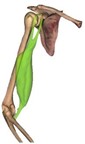
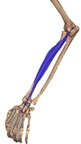
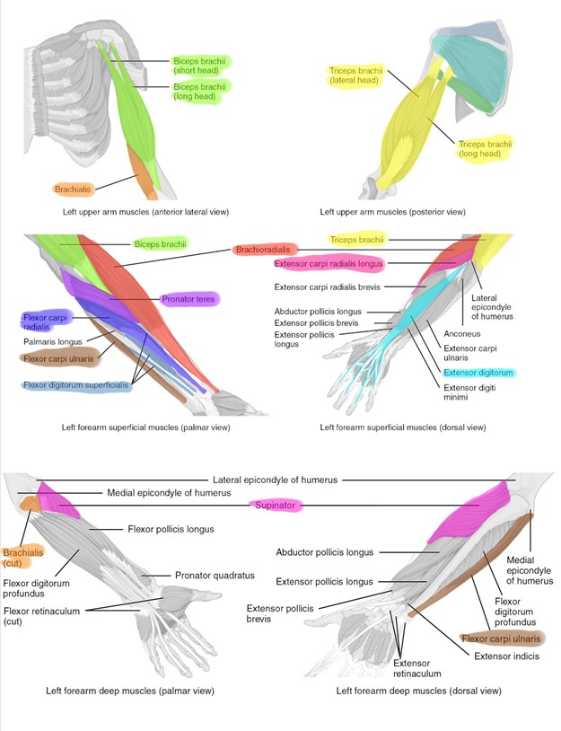
Note that, when in anatomical position, most of the muscles that act as flexors are found on the anterior surface or the arm and conversely, the muscles that act as extensors are typically found on the posterior surface of the arm. The forearm flexors are the biceps brachii, brachialis, and brachioradialis. As the name implies, the biceps brachii has two heads. The brachialis is deep to the biceps brachii and is an agonist of elbow flexion. The main extensor is the triceps brachii which has three heads. Another assisting extensor muscle is the anconeus. The forearm can also pronate (when the forearm faces posteriorly) and supinate (when the forearm faces anteriorly). The prime pronator muscle is the pronator teres and the supinator muscle controls supination. These two muscles can be found crisscrossing over the radius and ulna to create the pivoting action.
Figure 8: Deep muscles of the shoulder from anterior lateral view (c) and posterior view (d).
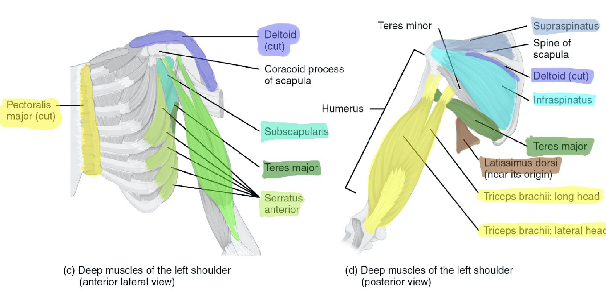
Table 9: Muscles that move the forearm and their action(s), origin(s), insertion(s) and innervation.
| Muscle | Image | Action | Origin | Insertion | Innervation |
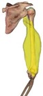
| Biceps brachii |
 |
supinates and flexes forearm | long head: supraglenoid tubercle, short head: coracoid process | radial tuberosity |
Musculocutaneous nerve |
| Brachialis | 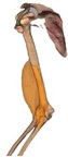 |
flexes forearm | distal anterior diaphysis of the humerus | tuberosity and coronoid process of ulna | Musculocutaneous nerve |
| Brachioradialis | 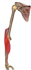 |
flexes forearm | ridge proximal to the lateral condyle of the distal humerus | styloid process of radius | Radial nerve |
| Pronator teres |
 |
pronates forearm | medial epicondyle of humerus, coronoid process of ulna | lateral surface of radius | Median nerve |
| Supinator | 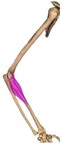 |
supinates forearm | lateral epicondyle of humerus, area of the radial notch of ulna | anterolateral area of proximal third of radius, distal to tuberosity | Radial nerve |
| Triceps brachii |
 |
extends and adducts arm | long head: infraglenoid tubercle of scapula; lateral and medial head: diaphysis of humerus | olecranon process of ulna | Radial nerve |
Table 10: Muscle actions of the forearm.
| Actions of the Forearm | Flexion | Extension | Pronation | Supination |
| Biceps brachii | X (PM) | X (PM) | ||
| Brachialis | X (PM) | |||
| Brachioradialis | X | |||
| Pronator teres | X (weak) | X | ||
| Triceps brachii | X (PM) | |||
| Supinator | X |
Muscles of the wrist (carpus) and hand (Figure 9, Tables 11 and 12)
The muscles of the forearm overlie the ulna and radius and serve to move the wrist, hand, and fingers. The wrist, hand, and fingers are capable of many complicated movements. Typically, the forearm muscles are the most difficult to find and learn because these muscles are in different layers and directions to assist in these numerous complex movements. When viewing the anterior forearm look for the most lateral, flat muscle called the brachioradialis. Medial to the brachioradialis are the flexor carpi radialis, palmaris longus, and the flexor carpi ulnaris muscles, in order. Deep to the palmaris longis is the flexor digitorum superficialis. In the posterior view, the extensor digitorum is an easy landmark as it had tendons running to digits 2-5. Medially from the extensor digitorum is the extensor carpi radialis longus and then back to the brachioradialis.
Table 11: Muscles that move the carpus (wrist) and hand and their action(s), origin(s), insertion(s) and innervation.
| Muscle | Image | Action | Origin | Insertion | Innervation |
| Extensor carpi radialis longus | 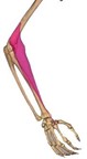 |
extends and abducts hand | lateral lower diaphysis of humerus | second metacarpal | Radial nerve |
| Extensor digitorum | extends carpus and hand |
lateral epicondyle of humerus |
posterior phalanges of fingers 2-5 |
Radial nerve |
|
| Flexor carpi radialis |
 |
flexes carpus and abducts hand |
medial epicondyle of humerus |
second and third metacarpals |
Median nerve |
| Flexor carpi ulnaris | 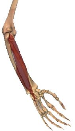 |
flexes hand; adducts hand |
medial epicondyle of humerus, coronoid process of ulna, anterior proximal radius | carpal bones, 5th metacarpal | Ulnar nerve |
| Flexor digitorum superficialis | 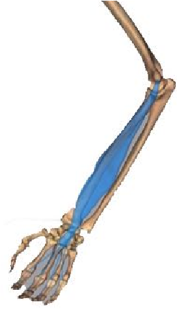 |
flexes wrist and flexes phalanges 2-5 | medial epicondyle of humerus, coronoid process of ulna, anterior proximal radius | middle phalanges of fingers 2-5 | Median nerve |
Table 12: Muscle actions of the wrist (carpus).
| Actions on the wrist | Flexion | Extension | Abduction | Adduction |
| Flexor carpi radialis | X (PM) | X | ||
| Flexor carpi ulnaris | X (PM) | X | ||
| Flexor digitorum superficialis |
X (PM) | |||
| Extensor digitorum | X (PM) | |||
| Extensor carpi radialis longus |
X | X |
Figure 9: Muscles of the forearm, wrist and hand.
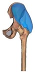
Muscles of the hip and thigh (femur) (Figures 10, 11, Tables 13, and 14)
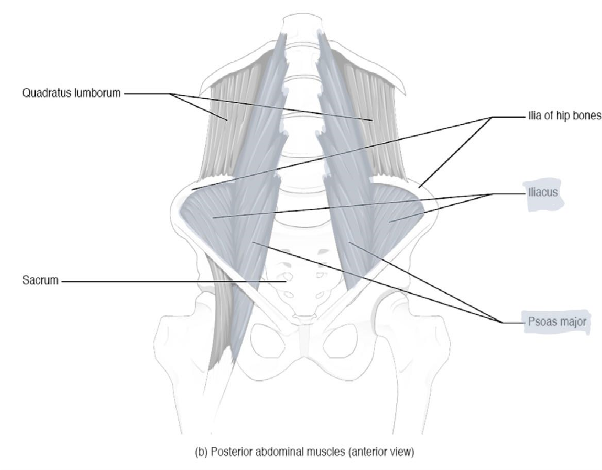
The anterior muscles of the hip and thigh flex the femur at the hip and extend the knee. This is an important action for the fore-swing phase during walking and for flexing the hip for sitting. The posterior muscles of the hip and thigh extend the thigh and flex the knee. This is the movement necessary for the backswing phase of walking and for standing. Muscles that flex the thigh originate from the vertebral column and pelvis and pass anteriorly to the hip joint. These muscles include the iliopsoas and rectus femoris. The iliopsoas muscle is formed from two muscles, the iliacus and psoas major. The posterior thigh muscles that extend the hip include the gluteus maximus and hamstring group, which itself consists of three muscles. As the name suggests, the gluteus maximus is a large muscle, located superficially on the posterior of the thigh.
Figure 10: Muscles of the hip, anterior view.
The lateral muscles of the thigh, like the middle-sized gluteus medius, control abduction (away from midline) of the hip. The medial muscles of the thigh cause adduction of the leg (bringing in toward midline). The three adductor muscles are the longus, brevis, and magnus. Their names suggest their shapes: the adductor longus is long and skinny, the brevis is the shortest (briefest), and the adductor magnus is the largest. The sartorius muscle runs obliquely across the upper thigh and its action is rotational. It makes a great landmark when examining the anterior thigh as it wraps laterally to medially and looks like a sash. The sartorius is the longest muscle in the body! The gracilis is a flat, strap-like muscle that can be seen on the medial thigh.
Table 13: Muscles of the hip and thigh and their action(s), origin(s), insertion(s) and innervation.
| Muscle | Image | Action | Origin | Insertion | Innervation |
| Adductor longus | adducts and flexes the thigh; rotates the thigh medially | body of pubis | linea aspera of femur | Obturator nerve | |
| Adductor magnus |
adducts and medially rotates the thigh | Ischial tuberosity, ischial ramus, inferior ramus of pubis | linea aspera and adductor tubercle of femur | Obturator and Sciatic nerves | |
| Gluteus maximus |
 |
extends and abducts the thigh; laterally rotates the thigh | posterior and lateral parts of the ilium, sacrum, and coccyx |
gluteal tuberosity of femur | Inferior gluteal nerve |
| Gluteus medius |
 |
abducts and medially rotates thigh | Outer surface of Ilium | greater trochanter of femur | Superior gluteal nerve |
| Gracilis | adducts and flexes the thigh, medially rotates thigh | body and inferior ramus of pubis | medial, proximal tibia | Obturator nerve | |
| Iliopsoas: Iliacus & Psoas major | 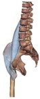 |
flexes thigh | Iliacus: iliac fossa Psoas major: lateral side and transverse processes of T12 and lumbar vertebrae | lesser trochanter of femur | Femoral nerve |
| Sartorius | flexes the thigh and leg (knee); abducts and laterally rotates the thigh | Anterior superior iliac spine | proximal, medial condyle of the tibia | Femoral nerve |
Table 14: Muscle actions of the hip and thigh.
| Actions at the hip and thigh | Flexion | Extension | Abduction | Adduction | Medial Rotation | Lateral Rotation |
| Iliacus | X (PM) | |||||
| Psoas major | X (PM) | |||||
| Adductor longus | X | X | X | |||
| Rectus femoris | X | |||||
| Sartorius | X | X | X | |||
| Biceps femoris | X (PM) | X (weak) | ||||
| Gluteus maximus | X (PM) | X | X | |||
| Adductor magnus | X | X | X | |||
| Semimembranosus | X | |||||
| Semitendinosus | X | |||||
| Gluteus medius | X (PM) | X | ||||
| Gracilis | X | X |
Figure 11: Muscles of the hip and thigh showing the superficial anterior (top), deep pelvic, anterior view (bottom, left) and posterior (bottom, right) views.

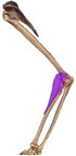
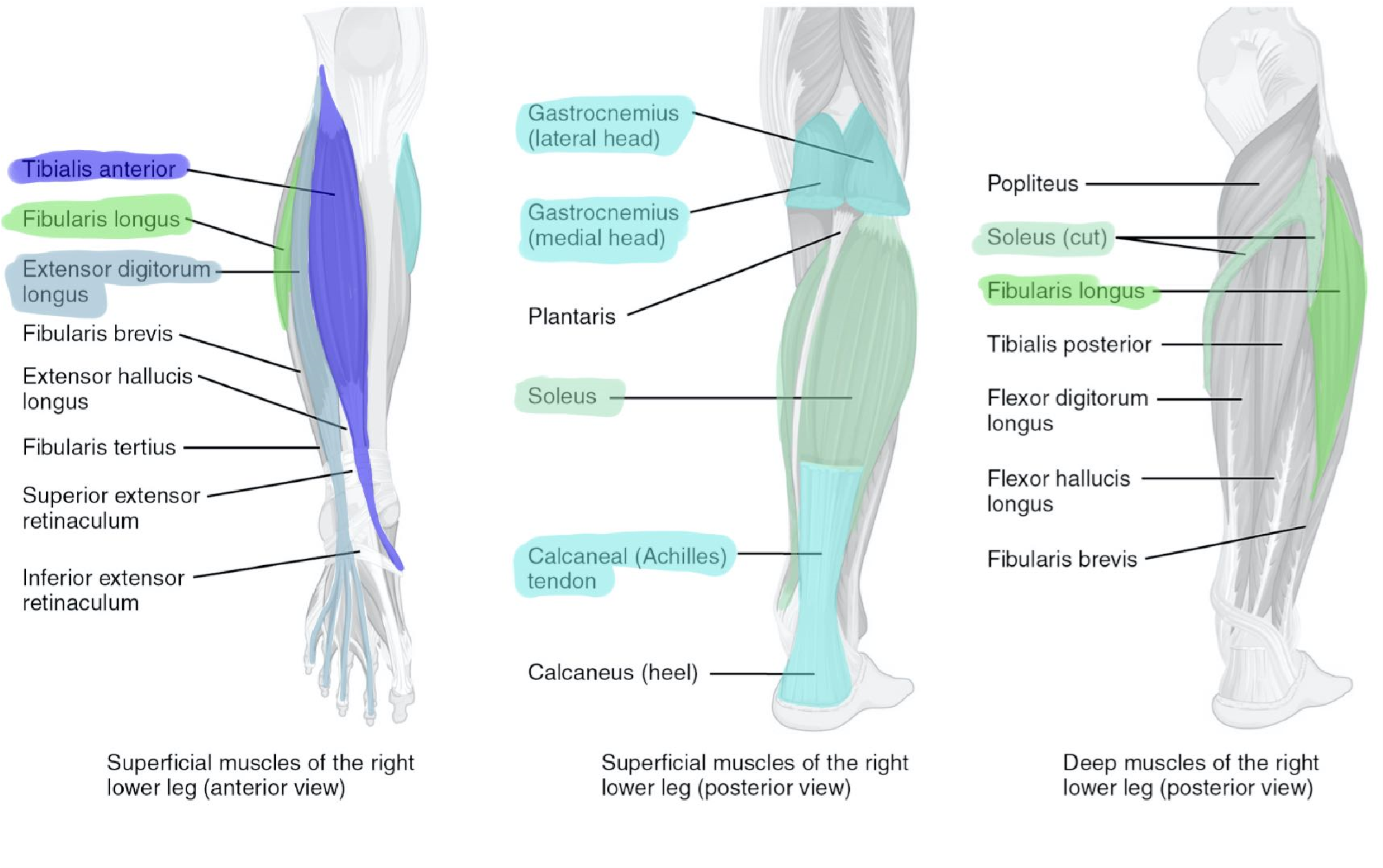
Muscles of the leg (tibia and fibula) (Figures 11, 12, Tables 15, and 16)
The muscles of the thigh extend and flex the lower leg at the knee. The quadriceps femoris is a group of four muscles on the anterior thigh and are the prime movers for knee extension. The rectus femoris is the middle muscle that is in the femoral region and connects to the patella. Lateral to the rectus femoris is the vastus lateralis. Medial to the rectus femoris is the vastus medialis, and deep to the rectus femoris is the vastus intermedius. On the posterior surface of the upper leg (femur) you can find the hamstring group. The hamstrings are a group of three muscles that are prime movers for knee flexion. The most lateral of the three muscles is the biceps femoris with two heads. The semimembranosus is the most medial muscle and on top of semimembranosus is the semitendinosus with the longest tendon.
Hint: Semimembranosus is medial and Semitendinosus is on top and has a long tendon.
Table 15: Muscles of the leg and their action(s), origin(s), insertion(s) and
innervation.
| Muscle | Image | Action | Origin | Insertion | Innervation |
| Biceps femoris |
flexes the leg (knee); laterally rotates and extends the thigh | ischial tuberosity (long head); linea aspera, and distal posterior femur (short head) | head of fibula and lateral condyle of tibia | Sciatic nerve | |
| Rectus femoris |
extends leg (knee), flexes thigh | anterior inferior iliac spine and acetabulum | patella and tibial tuberosity via quadriceps femoris tendon | Femoral nerve | |
| Semimembranosus | flexes the leg (knee); extends and medial rotates the thigh | ischial tuberosity | posterior surface of medial condyle of tibia | Sciatic nerve | |
| Semitendinosus | flexes the leg (knee); extends the thigh; medially rotates the leg | ischial tuberosity | proximal, medial surface of the tibia | Sciatic nerve | |
| Vastus intermedius |
extends leg (knee) | anterior and lateral part of the proximal diaphysis of the femur | quadriceps femoris tendon; lateral patella; lateral condyle of tibia | Femoral nerve | |
| Vastus lateralis |
extends leg (knee) | greater trochanter, proximal linea aspera of femur | quadriceps femoris tendon; lateral patella | Femoral nerve | |
| Vastus medias |
extends leg (knee) | linea aspera of femur | medial border of patella | Femoral nerve |
Table 16: Muscle actions on the lower leg.
| Actions on the leg (knee) | Flexion | Extension |
| Rectus femoris | X (PM) | |
| Vastus intermedius | X (PM) | |
| Vastus lateralis | X (PM) | |
| Vastus medialis | X (PM) | |
| Biceps femoris | X (PM) | |
| Semimembranosus | X (PM) | |
| Semitendinosus | X (PM) | |
| Gastrocnemius | X | |
| Gracilis | X | |
| Sartorius | X |
Muscles of the ankle and foot (Figure 12, Tables 17, and 18)
Muscles acting on the ankle joint cause dorsiflexion or plantarflexion. Muscles acting on the intertarsal joints allow for eversion and inversion. The muscles are found overlying the tibia and fibula of the lower leg. The anterior tibia is an excellent landmark for the lower leg muscles. Just lateral to the anterior surface of the tibia is the tibialis anterior, literally “front of the tibia”. Moving laterally from the tibialis anterior is the extensor digitorum longus and the most lateral muscle on the fibula is the fibularis longus (peroneus longus). Muscles on the anterior aspect of the tibia are responsible for extending the toes and dorsiflexing the foot. Muscles on the lateral aspect of the leg (fibula) are responsible for eversion and plantarflexing the foot. The gastrocnemius is the large calf muscle found on the posterior leg with the soleus deep to it. The gastrocnemius forms the calcaneal tendon (Achilles’ heel) that inserts onto the tarsal bone, the calcaneus. This posterior muscle performs one of the most powerful actions, plantar flexion. Plantar flexion allows humans to walk and run by providing forward thrust. The Soleus is a flat muscle deep to gastrocnemius; it assists in plantar flexion.
Table 17: Muscles of the ankle and foot and their action(s), origin(s), insertion(s),
and innervation.
| Muscle | Image | Action | Origin | Insertion | Innervation |
| Extensor digitorum longus | dorsiflexion of the foot and extends the toes | lateral condyle of tibial and proximal fibula | 2-5 middle phalanges | Deep fibular nerve | |
| Fibularis longus (peroneus longus) | eversion and plantarflexion of the foot | proximal lateral fibula | medial cuneiform bone and first metatarsal | Superficial fibular nerve | |
| Gastrocnemius | flexes the knee and plantarflexes the foot | medial and lateral condyles of femur | posterior calcaneus via calcaneal tendon | Tibial nerve | |
| Soleus | plantarflexes the foot | head of fibula, proximal tibia | posterior calcaneus | Tibial nerve | |
| Tibalis anterior | dorsiflexion and inverts the foot | lateral condyle and proximal 2/3 of tibial diaphysis | medial tarsal bone (cuneiform) and first metatarsal bone | Deep fibular nerve |
Table 18: Muscle actions on the ankle.
| Actions on the foot (ankle) |
Plantar Flexion |
Dorsiflexion | Inversion | Eversion |
| Gastrocnemius | X (PM) | |||
| Soleus | X (PM) | |||
| Fibularis longus (Peroneus longus) | X | X | ||
| Tibialis anterior | X (PM) | X | ||
| Extensor digitorum longus | X |
Figure 12: Muscles of the lower leg. Superior muscles, anterior view (left), superior muscles, posterior view (middle) and deep muscles, posterior view (right).
Procedure:
1. Divide into pairs. One pair will label and answer the questions on the arm muscle model while the other pair works on the leg muscle model. As you identify the muscles, label them with the laminated muscle stickers. Fill in the charts with the muscle name that matches the number on the model and draw a line between the muscle name and the muscle on the images.
2. Switch models and repeat step 1.
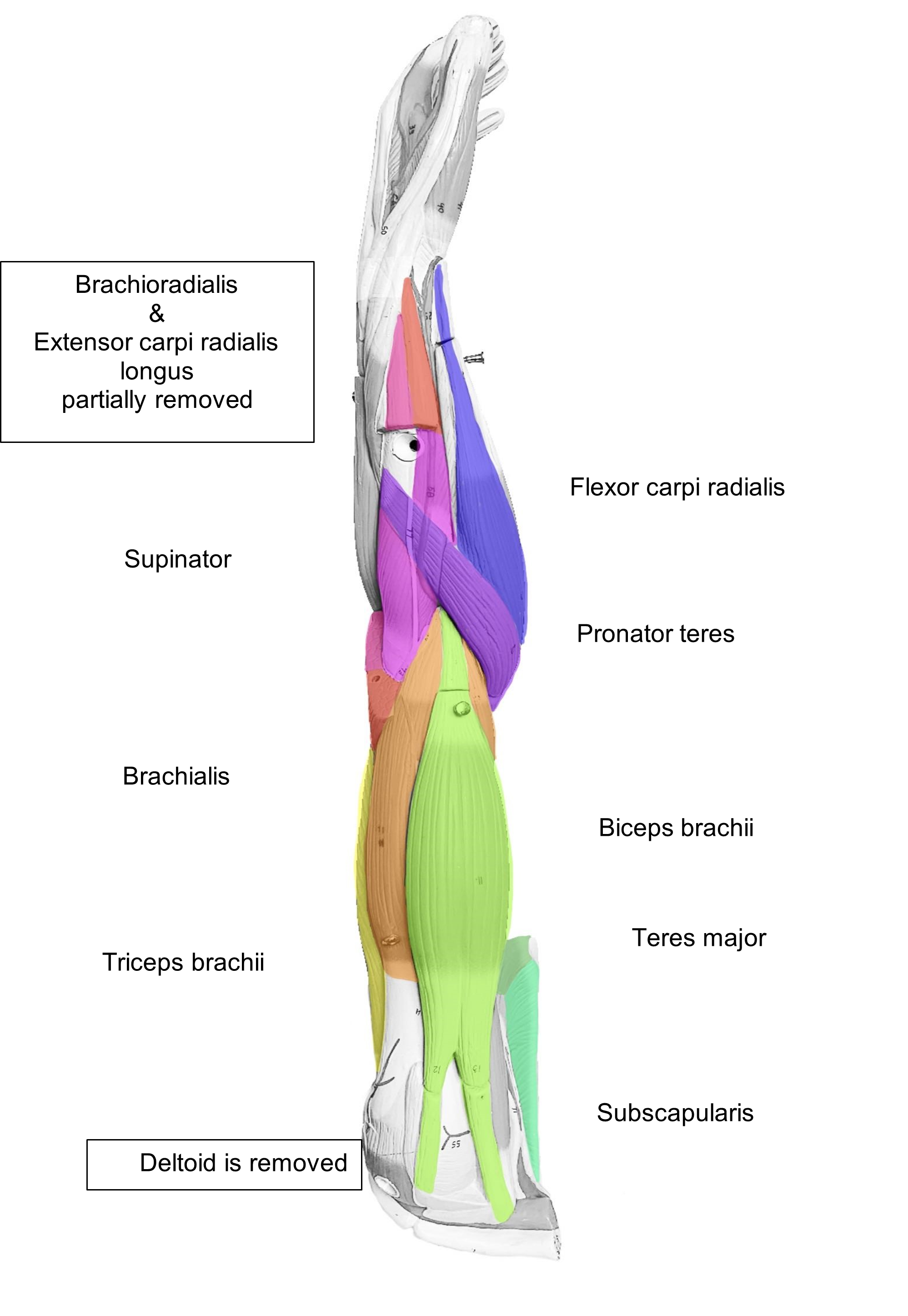
| Number | Muscles of the Arm |
| 5 | |
| 6 | |
| 7 | |
| 9 | |
| 10 | |
| 11, 12, & 13 | |
| 14 | |
| 16, 17, & 18 | |
| 19 | |
| 20 | |
| 22 | |
| 23 | |
| 25 | |
| 26 | |
| 30 | |
| 34 |
Word Bank: Biceps brachii, Brachialis, Brachioradialis, Deltoid, Extensor carpi radialis longus, Extensor digitorum, Flexor carpi radialis, Flexor carpi ulnaris, Flexor digitorum superficialis, Infraspinatus, Pronator teres, Subscapularis, Supinator, Supraspinatus, Teres major, Triceps brachii
Anterior Arm Muscles to Label
Posterior Arm Muscles to Label
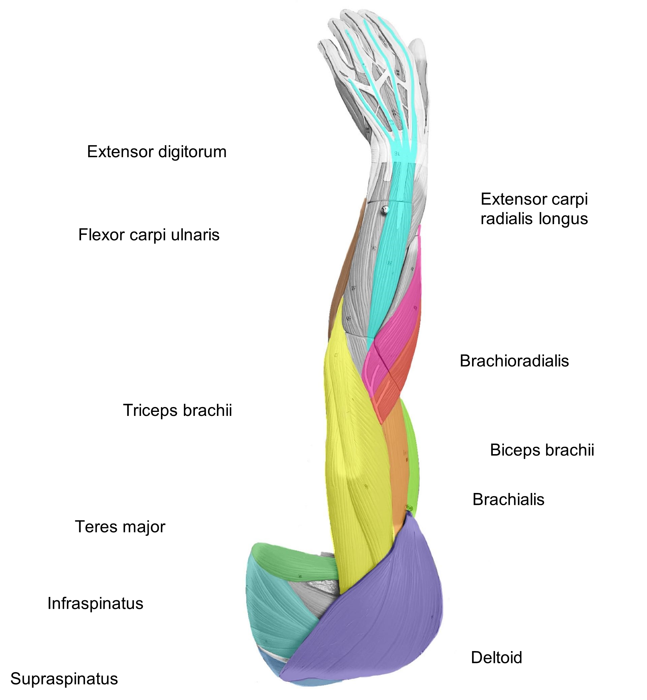
Lateral Arm Muscles to Label
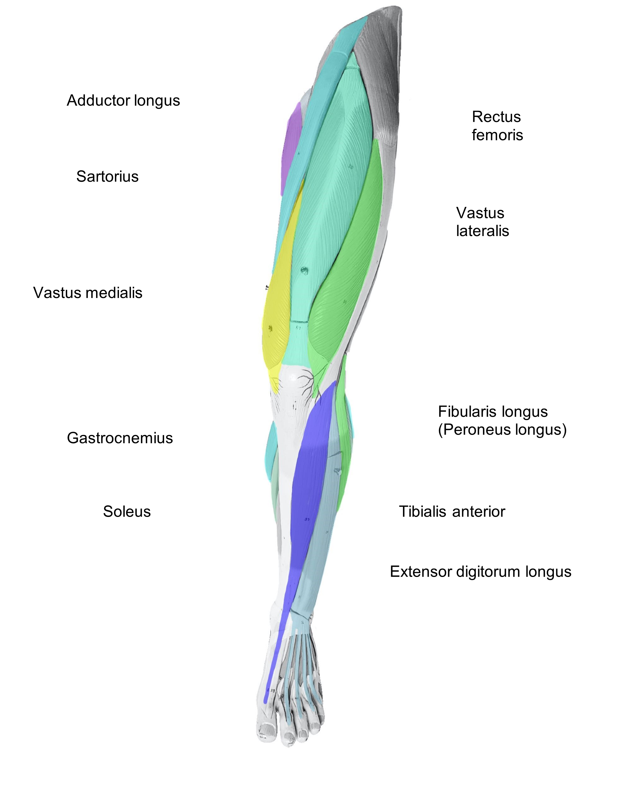
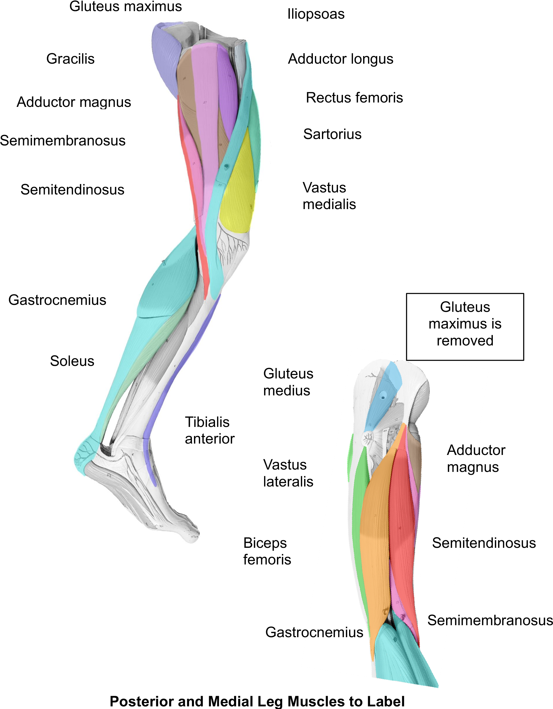
3. As you work on the leg model fill in the key using the numbers of the model and matching them to the numbers of your leg chart. Label the images of the leg model using the same key.
| Number | Muscles of the Arm |
| 11 | |
| 12 | |
| 20 | |
| 21 | |
| 22 | |
| 23 | |
| 24 | |
| 25 | |
| 26 | |
| 27 | |
| 28 | |
| 29 | |
| 30 | |
| 31 & 32 | |
| 33 | |
| 34 | |
| 35 | |
| 37 & 38 | |
| 39 |
Word Bank: Adductor longus, Adductor magnus, Biceps femoris, Extensor digitorum longus, Fibularis longus (Peroneus), Gastrocnemius, Gluteus maximus, Gluteus medias, Gracilis, Iliopsoas, Rectus femoris, Semimembranosus, Semitendinosus, Soleus, Tibialis anterior Vastus lateralis, Vastus intermedius, Vastus medialis, Sartorius
Anterior Leg Muscles to Label
[ ]
Posterior and Medial Leg Muscles to Label
4. Identify the muscles of the axial and appendicular on the full body muscle model. Draw a line from the muscle name to the muscle on the image.
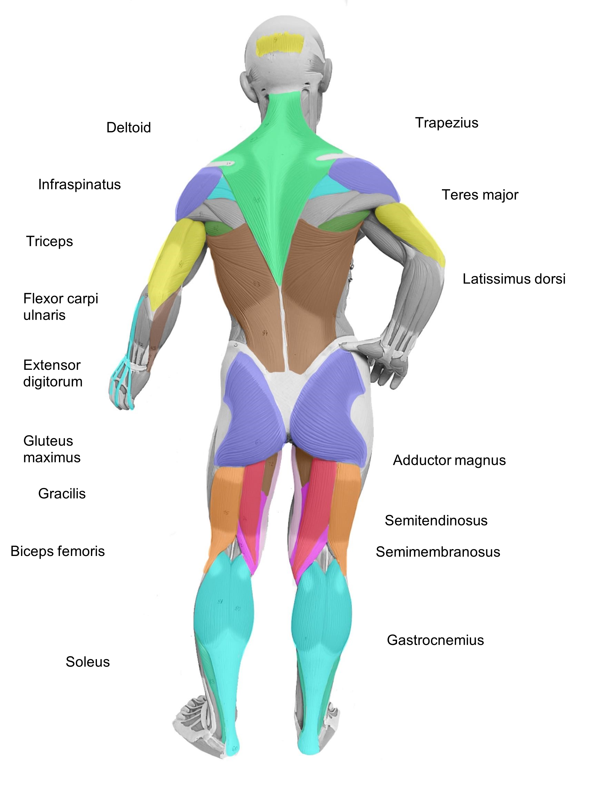
Posterior Body Muscles to Label
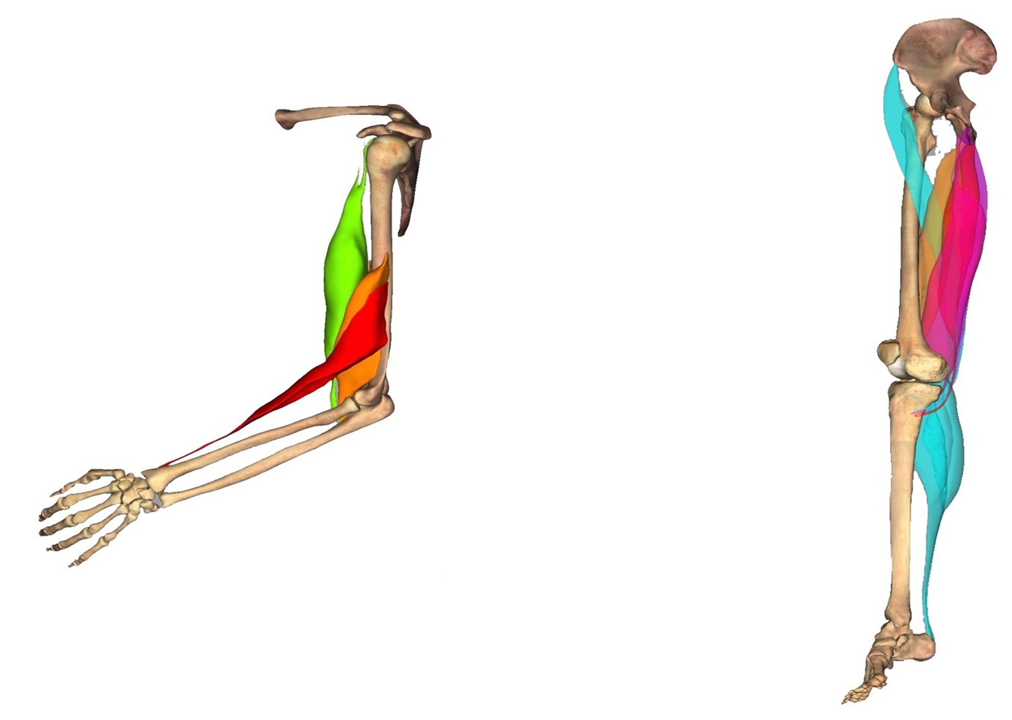
5. Look at the following two images. Draw with an arrow the direction of contraction of each muscle group. Name the angular movement indicated by each arrow.
Angular Movement
6. Once you have labelled all the images in the lab, ask your instructor to check and initial below.
Instructor’s Initials for labeled muscle: _____________________
Putting It All Together
Your guess was correct, the rectus femoris was the muscle on the anterior thigh. You listen as the doctor talks to the family about the severity of the disease. As of today, there is no cure for Duchenne; it is an irreversible, progressive disease. Jose will undergo intense physical therapy to keep his muscles active and moving for as long as possible. Eventually, the muscles of respiration will weaken and cannot move air into the lungs. Jose will receive breathing support at this point. In addition to skeletal muscle weakness, Duchenne also affects the heart (cardiac) muscle. Thus, heart contractions cannot keep up with the demands of the body. This is a sad diagnosis for the family. This case highlights how the muscles in the body work together in synchrony to produce incredible movements that support the body and life itself.
Creative Commons Citations:
- Muscles Lab, Case-Study Based OER Lab Manual for A&P I © 2022 by G. Backus, H. Wangerin, P. Rodgers is licensed under CC BY-NC-SA 4.0.
- Joint Movements, Anterior Body Muscles, Posterior Body Muscles, Anterior Arm Muscles to Label, Posterior Arm Muscles to Label, Lateral Arm Muscles to Label, Anterior Leg Muscles to Label, Posterior and Medial Leg Muscles to Label, Posterior Body Muscles to Label, © 2022 by H. Wangerin is licensed under CC BY-NC-SA 4.0.
- Muscle Action Predictions, Angular Movement, Muscle Actions, Posterior View of Leg, and images in Tables 2, 3, 4, 5, 6, 7, 9, 11, 13, 15, 17 and © 2022 by H. Wangerin, using the Anatomage Table, is licensed under CC BY-NC-SA 4.0.
- Figures 1, 2, 3, 4, 5, 6, 7, 8, 9, 10 11, and 12, Head Muscles to Label Abdominal Muscles to Label, © 2023 OpenStax Anatomy and Physiology 2e is licensed under CC BY 4.0 with color shading added by H. Wangerin.
