
23 Post-Lab for The Appendicular Skeleton
Gillian Backus; Heidi W. Wangerin; and Paula Rodgers
Post-Lab Activities: The Appendicular Skeleton Lab
Name: ________________________
Lab Checkout:
When you finish the lab, please clean up your lab space and put away your materials neatly in the tray. Once you have thoroughly cleaned, washed, and dried your lab table, please get your instructor’s initials to check-out of lab.
- Lab bench clean, washed, and dried
- Materials put away properly and organized in trays
- Microscope properly put away
Lab completed (% completed = ______ %) Instructor initials: ______________
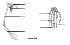
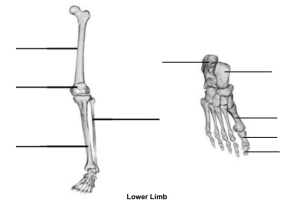
Activity 1: Bones of the Appendicular Skeleton
1. Label the bones of the upper limb, lower limb, foot, and hand. Give specific bone name(s) ie. metatarsal I or distal phalanx II.
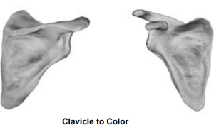
Activity 2: Bone Markings of the Upper Limb
2. Color the bullet point (or term) and the corresponding surface of the bone marking on the scapula in the same color:
-
- Acromion
- Coracoid process
- Glenoid cavity
- Infraspinous fossa
- Spine
- Subscapular fossa
- Supraspinous fossa
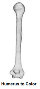
3. Color the bullet point (or term) and the corresponding surface of the bone marking on the humerus in the same color.
-
- Capitulum (lateral condyle)
- Deltoid tuberosity
- Greater tubercle
- Head
- Neck
- Olecranon fossa
- Trochlea (medial condyle)
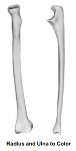
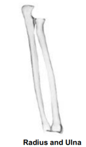
4. Color the bullet point (or term) and the corresponding surface of the bone marking on the radius and ulna in the same color:
Radius
-
- Head
- Radial styloid process
- Radial tuberosity
- Ulnar notch
Ulna
-
- Coronoid process
- Head
- Olecranon process
- Radial notch
- Trochlear notch
- Ulnar styloid process
Activity 3: Bone Markings of the Lower Limb
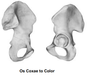
5. Label the following bone markings on the Os Coxae:
Ilium
- Acetabulum
- Anterior inferior iliac spine
- Greater sciatic notch
- Iliac crest
Ischium
- Ischial spine
- Ischial tuberosity
Pubic bone
- Obturator foramen
- Pubic symphysis
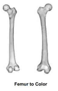
6. Color the bullet point (or term) and the corresponding surface of the bone marking on the femur (below) in the same color: Be sure to label which bone indicates the anterior view and which the posterior view.
- Fovea capitus
- Greater trochanter
- Head
- Lateral condyle
- Lateral epicondyles
- Lesser trochanter
- Medial condyle
- Medial epicondyles
- Neck
- Patellar surface
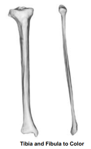
7. Color the bullet point (or term) and the corresponding surface of the bone marking on the tibia and fibula (below) in the same color:
Tibia
- Lateral condyle
- Medial condyle
- Medial malleolus
- Tibial tuberosity
Fibula
- Head
- Lateral malleolus
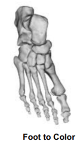
8. Color the bullet point (or term) and the corresponding bone(s) on the tarsus (ankle) and foot in the same color:
Ankle
- Calcaneus
- Talus
Foot
- Metatarsal I
- Metatarsal II
- Metatarsal III
- Metatarsal IV
- Metatarsal V
- Proximal phalanx
- Middle phalanx
- Distal phalanx
9. On the image below, circle the articulation points between the radius and ulna. Label the bone markings involved in the articulation points.
10. How many total phalanges are found in both hands and feet?
11. Read the following bone description and guess which bone is being described.
-
- You pick up a long, thin bone. One end has a sharp process, and the other end has a large “C” shaped notch. _____________________
12. Describe the following bones using at least three terms from your bone markings chart for each bone.
- Femur
- Tibia
- Scapula
